
Translate this page into:
Retrospective study of the pattern of eye disorders among patients in Delta State University Teaching Hospital, Oghara

-
Received: ,
Accepted: ,
How to cite this article: Ohwin PE, Abadom EG, Nwogueze BC, Nwabuoku US, Beteren IG. Retrospective study of the pattern of eye disorders among patients in Delta State University Teaching Hospital, Oghara. Calabar J Health Sci 2023;7:29-34.
Abstract
Objectives:
Ocular disease is an important public health issue and constitutes one of the commonest problems presenting in general primary healthcare in Nigeria. The present study examined the prevalence of eye disorders among patients attending Delta State University Teaching Hospital Oghara Delta State, Nigeria.
Material and Methods:
A descriptive study was adopted as the research design in sampling 2,003 cases in the hospital records within one (1) year using a simple random sampling technique.
Results:
Data obtained from the study revealed that 1033 (52%) of the cases reviewed were males, while 970 (48%) were females. The cases that were between the ages of 60–69 years were 395 (19.7%) out of which 216 (8.9%) were males and the remaining 179 (8.9%) cases were females. Majority of the cases reviewed were married with a frequency of 1,399 (69.9%). The occupational distribution of patients revealed that the highest ocular disease was found among civil servants with a total of 480 (24%) cases. Results of the study equally revealed that eye disorders related to the crystalline lens were the most prevalent eye diseases accounting for 620 (31%) out of the total sampled cases with cataract being the most prevalent (455, 22.7%) in this group. Conjunctiva eye disorders accounted for 320 (16%), followed by refractive errors 292 (14.6%), and retina diseases 262 (13.1%). Glaucomatous eye diseases were the fifth most detected with a total of 230 (11.5%). More so, the prevalence of optic nerve and visual pathway diseases was 86 (4.3%) out of the recorded cases while Patients with Eyelids disease had the lowest detected eye diseases with a total of 59 (3%), whereas, eye diseases of the Cornea seen was low 132 (6.6%).
Conclusion:
Eye conditions such as cataracts, conjunctivitis, refractive errors, glaucoma, cornea disorders, optic nerve, and visual pathway diseases, and eyelid disorders such as chalazion, blepharitis, and lipoma were identified to have a leading rate of a pattern of distribution with chances of complications. Hence, it is recommended that there is an urgent need for follow-up of patients of eye disorders with proper education on eye management to improve awareness of the pattern of eye disorder that could be associated with burden of visual impairment and blindness.
Keywords
Ocular diseases
Cataract
Glaucoma
Cornea disorder
Conjunctivitis
Refractive error

INTRODUCTION
Ocular diseases are important public health issues and constitute one of the commonest problems presented to the general practice clinic (10–21%) and could have significant socioeconomic consequences ;[1] as well as educational challenges, but also reduce the quality of life and increase the risk of death.[2] The incidence of various eye diseases in a community or area varies with the social and environmental factors. According to the Global Burden of Diseases,[3] previous studies have reported that worldwide pre disposable factors causing injuries and other health risk factors occurs among 41.9 million people due to legal blindness, 253.08 million people with moderate vision impairment, and 33.78 million people with severe vision impairment. Data from the World Health Organization showed that at least 2.2 billion people were affected by blindness and vision impairment around the world, of whom 1 billion had preventable vision impairment or impairment that had yet to be addressed.[2]
The pattern of occurrence of ocular diseases globally is dependent on factors such as age, gender, region, and climatic conditions.[4] The majority of these ocular conditions which can lead to blindness are either potentially preventable or curable.[5] Disease pattern is in a state of constant flux, over some time. The pattern of eye diseases differs in developing and developed countries and often in communities.[6] A study of the pattern of ocular diseases is very important because while some eye conditions are just causes of ocular morbidity others may lead to blindness.[5] Uncorrected refractive error, cataracts, age-related macular degeneration, glaucoma, conjunctivitis, corneal ulcers, uveitis, pterygium, and diabetic retinopathy are the leading causes of vision impairment globally.[7]
Eye diseases linked to blindness have remained a genuine issue that has a colossal and wide effect on the general public, with financial misfortune as it is usually accompanied by challenges in physical capacity, passionate misery, low socialization mental, and social wellbeing. The negative impact extends from the immediate family to society at large. Studies of ocular diseases are important in understanding the causes of ocular morbidity that could lead to visual loss.[1] Considering the complicated epidemiology of visual impairment and the wide variety of factors involved, region-specific intervention strategies are required for every community. Hence, this study evaluated the pattern of eye disorders among patients seen in Delta State University Teaching Hospital, Oghara, Delta State.
MATERIAL AND METHODS
Study design
This was a retrospective descriptive study, on the pattern of eye disorders among patients visiting Delta State University Teaching Hospital Oghara, Delta state, Nigeria.
Study area
The research study was carried out in the Opthalmology and Optometry Unit of Delta State University Teaching Hospital Oghara, Delta State Nigeria. Oghara is a town located in Ethiope West Local Government Area of Delta State, Nigeria. It is home to the Urhobo ethnic group of Delta State. The town is known for its rich mineral resources and their major occupations are mainly farming and trading. Oghara is a town with an average population of 150,000 people.
Sample and sampling technique
The study consisted of both male and female patient case files at the records unit of the eye clinic section of the Delta State University Teaching Hospital Oghara Delta state, Nigeria. The sample size for the study was 2003 case files available in the records department within 1 year.
Method of data collection
A well-structured data collection sheet was used to record the data collected from the file case from the medical record at the ophthalmology and optometry unit in Delta State University Teaching Hospital, Oghara Delta State separating the different types of eye diseases from June 2018 to May 2019. The data collection sheet included the following: age and gender of the subjects, occupation, marital status, and eye disorder diagnoses.
Inclusion and exclusion criteria
The eye clinic session of the Delta University Teaching hospital is known as Ophthalmology Department. It comprise of the ophthalmologist, optometrist and other eye health personnel. The selection criteria was based on information provided by the ophthalmology department record unit. Only General Out Patients (GOP) case files within study duration having records of patients’ sex, age, occupation, marital status as well as ocular diagnosis were recruited. Patients case files domicile in this record unit, having diagnosis which are not eye related disease condition were excluded.
Ethical consideration
Ethical approval was obtained from ethical committee of both the Delta State Teaching Hospital, Oghara where the data were collected and from the Human Physiology Department of Delta State University, Abraka where the research topic was approved and supervised as a BSc project work. The study was duly approved as a retrospective study requiring case files only.
Data analysis
The collected data were entered into SPSS version 23 for analysis. Descriptive analyses that include proportion, percentage, and appropriate graphics presentations were applied to describe the data.
RESULTS
The results on the pattern of eye disorders at Delta State University Teaching Hospital, Oghara are presented in [Figures 1-4 and Table 1]. A total of 2,003 patients were registered and examined in the hospital within the one (1) year period.

- Sex distribution of the patients seen.
- Age and sex distribution of the patients seen.

- Distribution of patients by marital status.

- Distribution of patients by occupation.
| Types of eye diseases | Frequency | Percentage |
|---|---|---|
| Glaucoma | 229 | 10.54 |
| Neovascular glaucoma | 48 | 2.40 |
| Primary open-angle | 126 | 6.30 |
| Primary angle closure | 20 | 1.00 |
| Norman tension glaucoma | 25 | 1.25 |
| Eyelids | 59 | 2.95 |
| Chalazion | 44 | 2.20 |
| Blepharitis | 9 | 0.45 |
| Lipoma | 6 | 0.30 |
| Retina | 273 | 13.65 |
| Diabetic retinopathy | 128 | 6.40 |
| Central retinal vein occlusion | 22 | 1.10 |
| Macular edema | 36 | 1.80 |
| Age-related macular degeneration | 48 | 2.40 |
| Retinal detachment | 28 | 1.40 |
| Hypertensive retinopathy | 11 | 0.55 |
| Optic nerve and visual pathway | 86 | 4.30 |
| Optic atrophy | 49 | 2.45 |
| Optic neuritis | 26 | 1.30 |
| Retinitis Pigmentosa | 11 | 0.55 |
| Crystalline lens | 620 | 30.98 |
| Unilateral cataract | 455 | 22.74 |
| Bilateral cataract | 142 | 7.10 |
| Subluxated cataract | 23 | 1.15 |
| Refractive errors | 292 | 14.59 |
| Myopia | 143 | 7.15 |
| Hypermetropia | 56 | 2.80 |
| Astigmatism | 93 | 4.65 |
| Cornea diseases | 132 | 6.60 |
| Corneal opacity | 79 | 3.95 |
| Keratitis | 30 | 1.50 |
| Corneal dystrophy | 23 | 1.15 |
| Conjunctiva | 320 | 15.99 |
| Allergic conjunctivitis | 204 | 10.19 |
| Bacteria conjunctivitis | 98 | 4.90 |
| Pterygium | 18 | 0.90 |
| Grand Total | 2,001 | 100.0 |
The sex of patients that were diagnosed with eye disease at the Delta state Teaching Hospital Oghara within study duration was found to be 52% (1033) male and 48% (970) female [Figure 1]. This implies that majority of patient that presented at the hospital for year 2003 and were diagnosed of eye disease were male patients. This also shows that female has less access to eye care.
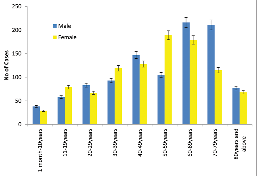
[Figure 2] above represents the age and sex distribution of cases that were reported to Delta State University Teaching Hospital, Oghara for diagnosis of eye diseases. Data obtained revealed that 67 (3.35%) cases reviewed for patients were between the ages of 1 month–10 years, 38 (1.9%) were male patients whereas, 29 (1.4%) cases were attributed to female patients. Patients between the ages of 11–19 years had a total of 137 (6.8%) of which males accounted for 58 (2.9%) and 79 (3.9%) were attributed to female cases. The prevalence of eye diseases among patients between the ages of 20–29 years revealed that a total of 150 (7.5%) cases were reviewed, out of which only 83 (4.1%) were male cases, while 67 (3.3%) were female cases. Similarly, a total of 212 (10.6%) cases were reviewed for patients between the ages 30–39 years, 93 (4.6%) were male patients whereas, 119 (5.9%) cases were attributed to female patients.
Additionally, Figure 2 showed that 275 (13.7%) of the cases reviewed were between ages of 40-49 years out of which 147 (7.3%) were male and 128 (6.4%) were female cases. The prevalence of eye diseases in this study for patients between the ages of 50-59 years were 294 (14.7%), out of which 105 (5.2%) were male cases and the other 189 (9.4%) were female cases. More so, 326 (16.3%) of cases reviewed were between the ages of 70-79 years, out of which 211 (10.5%) were male and 115 (6.4%) were female cases. The final category reviewed were between the ages of 80 years and above; it had a total of 145 (7.3%) cases reviewed, out of which 77 (3.8%) were male and the remaining 68 (3.4%) were female. Summarily, based on only sex distribution [Figure 1], a total of 2003 cases were reviewed out of which 1033 (52%) were male patients and the remaining 970 (48%) were female patients.
[Figure 3] represents the marital status of patients. A total of 390 (19.5%) cases reviewed were still single patients, whereas, 1399 (69.9%) cases were married, 191 (9.5%) were widows or widowers, and the remaining 21 (1%) were divorced. This implies that the majority of the cases diagnosed with eye disease in the study area were married.
Occupational distribution of patient [Figure 4] showed that 24% (480) of subjects were civil servants,19.2% (385) were traders, 18.1% (362) were farmers while the least distribution percentage were students 17.4% (349). However, patients with other occupational statuses accounted for 133 (6.6%) of the cases. This implies that more civil servants were diagnosed with eye disease in the study setting.
The pattern of various eye diseases in Delta State University Teaching Hospital, Oghara is shown in [Table 1]. Results of the study showed that eye diseases related to crystalline lenses were the most prevalent eye disorder accounting for 620 (31%) out of the total sampled cases. In this group, Unilateral Cataract was the most prevalent (455, 22.7%), followed by Bilateral cataract (142, 7.1%), and the least were Subluxated cataract (23, 1.2%). Patients with a diagnosis of Conjunctiva eye diseases were the second most detected eye diseases in the study area, with a total of 320 (16%). In this category, allergic conjunctivitis was the most prevalent (204, 10.1%), followed by bacteria conjunctivitis (98, 4.9%), and the least was Pterygium (18, 1%).
Results of the study showed that refractive errors were the third most detected group that result in visual impairment revealed with a total of 292 (14.6%). In this group, the most prevalent was myopia (143, 7.2%), this was immediately followed by patients diagnosed with hypermetropia (93, 4.7%), and the least was astigmatism (56, 2.8%). The prevalence of eye disease related to the Retina was the fourth most prevalent as recorded in this study with a total of 273 (13.6%) cases. Prevalence noted in this group were; diabetic retinopathy (128, 6.4%), age-related macular degeneration (48, 2.4%), macular edema (36, 1.8%), retinal detachment (28, 1.4%), central retinal vein occlusion (22, 1%), and ocular hypertension (11, 0.6%) had the lowest number of cases.
Cases of glaucoma eye diseases were the fifth most detected with a total of 229 (10.5%). In this category, primary open-angle (126, 6.3%) was the highest followed by advanced glaucoma (48, 2.4%), bilateral glaucoma (25, 1.3%), and primary angle closure (20, 1%) which is the least. Eye diseases of the Cornea were the sixth most detected with a total of 132 (6.6%). In this group, corneal ulcer (79, 4%) was the highest, followed by keratitis (30, 1.5%) and corneal opacity (23, 1.2%) was the least. More so, the prevalence of optic nerve and visual pathway diseases was the seventh most prevalent with a total of 86(4.3%) out of the recorded cases. In this group Optic atrophy (49, 2.5%) was the highest, followed by optic neuritis (26, 1.3%) and hemianopia (11, 0.6%) were the least recorded cases. Patients with Eyelids disorder had the lowest prevalence with a total of 59 (3%). In this group, chalazion accounted for 44 (2.2%) as the highest case, this was followed by blepharitis, (9, 0.5%), while Lipoma (6, 0.3%) was the least observed.
DISCUSSION
The results gathered revealed that 1033 (52%) of the cases reviewed were males, whereas, while 970 (48%) were females. This implies that the majority of the cases of eye disease prevalent in the study area were males. This study is similar to the general belief that more males are seen in the clinics than females.[8] The finding from this study is similar to that of Bhoi,[1] where out of 2348 patients included in the study, the majority 1364 (58.1%) were male and 984 (41.9%) were females. The finding in our study was contrary to the study of Achigbu et al.,[9] where most of the study group found were female. Ademe and Edmealem[10] in their study ascertained that 197 (51.3%) of them were females and 186 (48.4%) were males. The study conducted on ocular morbidity in rural Ethiopia indicated that, a total of 214 patients were examined, where males comprised 50.5%.[11] This might be due to differences in sample size and gender patterns of countries.
Our study revealed that majority of the cases reviewed on eye diseases were prevalent among the older male patients between the ages of 60-69 years. The study from Muhammad and Dantani in Sokoto State Nigeria is comparable to our study in terms of the study subjects (all ages). In the study of Baranwal et al.,[12] the majority of age distribution was 51– 65 years (17.91%) in their study subject.
Worldwide, the pattern of ocular diseases varies from one location to another. However, cataracts, glaucoma, conjunctivitis, corneal ulcers, uveitis, refractive errors, and pterygium are considered common ocular disorders.[8] Cataracts, refractive errors/low vision, trachoma, onchocerciasis, and Vitamin A deficiency/other causes of childhood blindness were determined to be responsible for 75% of all blindness worldwide. In this present study the pattern of various eye diseases in patients seen at Delta State University Teaching Hospital, Oghara showed that ocular diseases related to the crystalline lens were the most prevalent eye disease. In this group, unilateral cataract was the most prevalent followed by bilateral cataract and the least were subluxated cataract. This finding is similar to a previous study by Monsudi et al.[13] Moreover, Bhoi[1] reported that Cataract was the third most common ocular morbidity.
Patients with a diagnosis of conjunctiva eye disorders were the second most detected eye diseases in the study area. In this category, allergic conjunctivitis was the most prevalent followed by bacteria conjunctivitis and the least was Pterygium Similarly, a study by Hassan et al., in South-West Nigeria noted that vernal conjunctivitis (21.1%) was the commonest disorder seen. On the contrary, Agyemang-Mireku[6] in Ghana found the commonest eye disorder to be conjunctivitis (39.70%). Similarly, a retrospective study in India showed that conjunctivitis was the commonest ocular disorder at 21.94%; others were cataract - at 9.2%, refractive error -at 15.2%, dacryocystitis - at 6.51%, and blepharitis - 3.2%.
Results of the study showed that refractive errors were the third most detected group of eye diseases seen. It was reported in the study of Baranwal et al.[12] that refractive error was the commonest ocular morbidity in their study followed by cataracts with follow-ups of cataract surgeries and allergic conjunctivitis which is contrary to this study. This study is in line with the study of Lakho and Ali,[5] who reported that Refractive error is the third most common ocular disease. This is in agreement with previous reports.[14] Balarabe et al.,[15] reported that refractive errors have a serious effect as it impacts the quality of life and consequently affect socioeconomic and educational life. In this group, the most prevalent was myopia, which was immediately followed by patients diagnosed with astigmatism and the least was hypermetropia which is contrary to observations by other authors Baranwal et al.[12] who reported that presbyopia was the most common refractive disorder in their study.
The prevalence of eye disease related to the Retina was the fourth most prevalent as recorded in this study. This is incomparable to what was found in the study of Adeoti et al.,[16] and Eze et al.[17] who reported that retina disease accounted for 111 (4.6%) and (3.9%) of cases in their respective studies. The prevalence noted in this group was diabetic retinopathy, age-related macular degeneration, macular edema, retinal detachment, and central retinal vein occlusion (22, 1%). This study is similar to the study of Nigeria and Malaysia and diabetic retinopathy accounted for 9.7% and 9.6% of retinal diseases, respectively.[5] Cases of Glaucoma eye diseases were the fifth most detected. This is similar to the report in tropical Africa where it came third after cataracts and aphakia.[16] However, it is not similar to the study of Glaucoma which came third contrary to previous reports where it has been reported as the second most common cause of visual impairment and blindness after cataracts.[8]
Eye diseases of the Cornea were the sixth most detected. In the study of Das et al.,[18] the second most common presentation was cornea and anterior segment disorders in Liberia which is different from this study. Similarly, in the study conducted by Bhoi[1] while in the study of Sharifi and Samadi,[19] on the pattern and prevalence of cornea disorders %). In our study, optic nerve and visual pathway diseases were the seventh most prevalent cases with optic atrophy being the highest, followed by optic neuritis and retinitis pigmentosa being the least recorded cases. Patients with Eyelid disorders had the lowest prevalence with chalazion being the highest case followed by blepharitis, while lipoma was the least observed.
CONCLUSION
From the study, eye conditions such as Cataracts, Conjunctiva, Refractive errors, Retina diseases, Glaucoma, Cornea disorder, Optic nerve, and visual pathway diseases, and Eyelid disorder such as chalazion, blepharitis, and Lipoma were identified. A cataract is the most common eye disease in this study population as the majority of them were male. It was observed that 395 (19.7%) of the cases reviewed were between the ages of 60–69 years. More attention should be paid to the management of these disorders through subspecialty development to reduce the burden of visual impairment and blindness in our environment and should be encouraged.
Declaration of patient consent
Patient consent not required as patients identity is not disclosed or compromised.
Conflicts of interest
There are no conflicts of interest.
Financial support and sponsorship
Nil.
References
- Pattern of ocular diseases in patients attending to a tertiary eye care center in Southern Odisha. IOSR J Dent Med Sci. 2018;17:1-3.
- [Google Scholar]
- Prevalence and causes of vision loss in China from 1990 to 2019: Findings from the global burden of disease study 2019. Lancet Public Health. 2020;5:e682-91.
- [CrossRef] [PubMed] [Google Scholar]
- Blindness and Vision Impairment Collaborators; Vision Loss Expert Group of the Global Burden of Disease Study. Causes of blindness and vision impairment in 2020 and trends over 30 years, and prevalence of avoidable blindness in relation to VISION 2020: The Right to Sight: An analysis for the Global Burden of Disease Study. Lancet Glob Health. 2021;9:e144-e160.
- [Google Scholar]
- Quantitative assessment of age and gender related changes in human lacrimal fluid composition in subjects. J Pharm Res Int. 2011;33:89-99.
- [Google Scholar]
- Pattern of eye diseases at tertiary eye hospital in Sudan (Makah Eye Hospital, Khartoum) Albasar Int J Ophthalmol. 2015;3:15-8.
- [CrossRef] [Google Scholar]
- Pattern of ocular conditions among patients attending an Eye Clinic in Ghana. Optometry. 2017;2:2476-5.
- [Google Scholar]
- Blindness and Vision Impairment. 2021. Data Sheet. Geneva: World Health Organization; Available from: https://www.who.int/news-room/fact-sheets/detail/blindness-and-visual-impairment [Last accessed on 2022 May 20]
- [Google Scholar]
- Pattern of ocular morbidity in Nigeria. Asian Pac J Trop Dis. 2013;3:164-6.
- [CrossRef] [Google Scholar]
- Ocular morbidity in rural communities in Imo State South East Nigeria. Open J Ophthalmol. 2016;6:184-90.
- [CrossRef] [Google Scholar]
- Pattern of ocular diseases among patients attending ophthalmic outpatient department: A cross-sectional study. Int J Clin Exp Ophthalmol. 2020;4:49-53.
- [CrossRef] [Google Scholar]
- Pattern of ocular diseases in children attending outpatient department of a rural medical college in central India. Int JSS. 2015;3:57-60.
- [Google Scholar]
- The prevalence of various eye diseases among patients of different nationalities attending the ophthalmology clinic at a tertiary care United Nations hospital: A 5 year retrospective analysis. Int J Contemp Med Res. 2019;6:I7-10.
- [CrossRef] [Google Scholar]
- Pattern of eye diseases present at a free outreach in a rural community in Northwestern Nigeria. Sudan Med Monit. 2015;10:113116.
- [CrossRef] [Google Scholar]
- Common ocular problems in Aba metropolis of Abia State, Eastern Nigeria. Pak J Soc Sci. 2009;6:32-5.
- [Google Scholar]
- Presbyopia among health workers in a tertiary hospital in North Western Nigeria. Sub Saharan Afr J Med. 2015;2:10-3.
- [CrossRef] [Google Scholar]
- Pattern of eye diseases in a tertiary hospital in Osogbo, Southwestern Nigeria. Asian J Res Rep Ophthalmol. 2020;3:1-8.
- [Google Scholar]
- The burden and spectrum of vitreo-retinal diseases among ophthalmic outpatients in a resource-deficient tertiary eye care setting in South-Eastern Nigeria. Middle East Afr J Ophthalmol. 2010;17:46-55.
- [CrossRef] [PubMed] [Google Scholar]
- Prevalence of ocular disorders in Liberia: A retrospective study using the eye Smart electronic medical record system. J Glob Health Rep. 2019;3:e2019033.
- [CrossRef] [Google Scholar]
- The frequency of ocular diseases in eye clinic at Imam Khomeini Hospital of Urmia. J Urmia Univ Med Sci. 2009;20:137-43.
- [Google Scholar]


